Research
Advantages and versatility of MALDI-TOF IMS for analyzing biological samples.
WHAT IS IMAGING MASS SPECTROMETRY (MS)?
In a broader context, imaging MS can be employed to investigate the molecular content of tissues for a wide range of applications from normal tissue, organ and body development, to pharmaceutical compound uptake in targeted organs and related toxic effects.
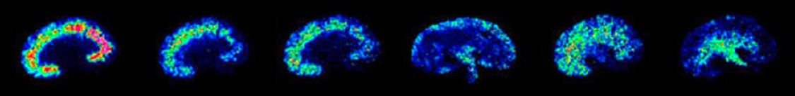
Imaging MS is the visualization of the localization of hundreds of different biocompounds present in thin tissue sections obtained by integrating the intensities of the signals observed throughout the course of data acquisition as a function of the tissue spatial coordinates.
The imaging MS technology can be used to query the molecular content of tissues for a wide range of applications. Examples of studies that can be conducted with this technology include:
- the study of disease-related biomarkers (portein, lipids...) in mammalian tissues;
- the expression of specific subsets of signals within recovered profiles and images that are linked with the resistance to existing therapies and, in some cases, patient outcome; and
- the molecular composition and organization in tissues from a wide range of organisms, including mollusks, insects and plants.
In a broader context, imaging MS can be employed to investigate the molecular content of tissues for a wide range of applications from normal tissue, organ and body development, to pharmaceutical compound uptake in targeted organs and related toxic effects.
As such, our lab collaborates with many reserachers in the biochemistry and medical domain. For concrete, published examples of research that we have contributed to, please refer to our publication page.
WHAT MS APPROACH DO WE USE?
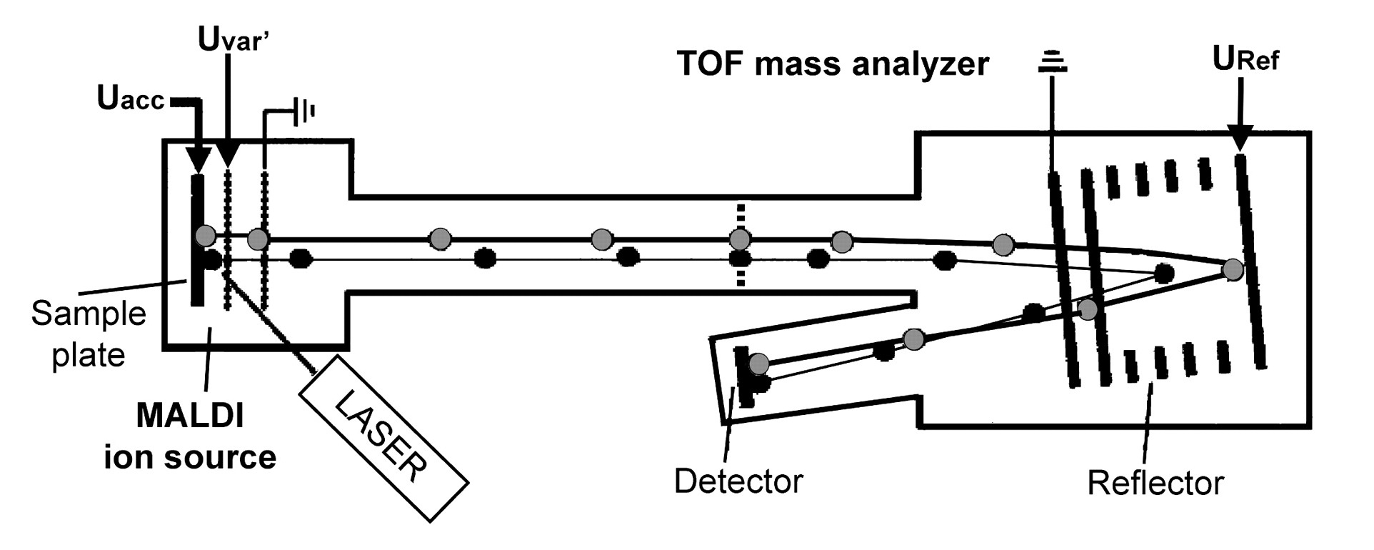
We primarily utilize matrix-assisted laser desorption ionization (MALDI) as means of ion production and time-of-flight (TOF) mass analyzers for ion detection. This approach allows us to analyze the proteomic content of both fresh frozen and formalin fixed paraffin embedded (FFPE) tisues, opening the possibility of investigating clinical samples stored for decades in tissue banks. You can find the model that we use on the instrumentation page.[/caption]
This content has been updated on 8 February 2022 at 16 h 44 min.